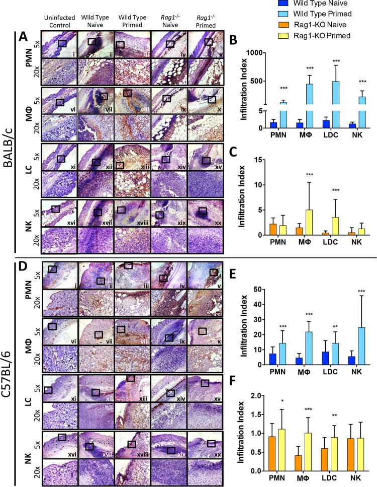
FIG 4.
Increased immune cells infiltrate into skin abscesses of primed and naive wild-type and rag1−/− mice. At day 7 postinfection, abscesses were dissected, fixed in zinc formalin, and embedded in paraffin. Skin sections from BALB/c (A) and C57BL/6 (D) mice were stained with antibodies against Ly6G for neutrophils (PMN) (i to v), F4/80 for macrophages (MΦ) (vi to x), CD207 for Langerin+ dendritic cells (LDC) (xi to xv), and CD49b for NK cells (NK) (xvi to xx). ImageJ software was used to quantify immune cell infiltration at abscess sites of wild-type (B and E) and rag1−/− (C and F) mice. *, P < 0.05; **, P < 0.01; ***, P < 0.001 (versus naive mice via Student's t test; n ≥ 10 images per sample). The infiltration index was calculated by determining the expression levels of samples (% of uninfected control levels), normalized to 106 CFU MRSA in abscesses of corresponding samples as previously described (53). Data presented are mean infiltration indices and SD.

