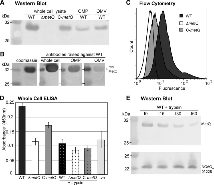
FIG 2.
Cell surface localization of MetQ. (A) Western blot of the N. gonorrhoeae 1291 wild type (WT), metQ knockout (ΔmetQ), and complemented (C-metQ) strains using polyclonal anti-MetQ antibodies. The samples analyzed included whole-cell lysates, OMP fractions, and OMVs. (B) Coomassie-stained SDS-polyacrylamide gel and Western blot of recombinant His-tagged MetQ (rec. MetQ) probed with polyclonal antibodies raised against either heat-inactivated whole cells, OMPs, or OMVs of the N. gonorrhoeae 1291 wild type. (C) Flow cytometry of whole cells of the N. gonorrhoeae wild type, ΔmetQ, and C-metQ strains, with the expression of MetQ on the cell surface being determined by the detection of binding of polyclonal anti-MetQ antibody and secondary Alexa Fluor 488 anti-mouse immunoglobulin antibody. (D) Whole-cell ELISAs of untreated and trypsin-treated (+trypsin) N. gonorrhoeae wild type, ΔmetQ, and C-metQ strains using polyclonal anti-MetQ antibodies. The results for the negative control (−ve), containing secondary antibody only, are also shown. The graph shows the average absorbance at 450 nm from three independent replicates ±1 standard deviation. By Student's t test, P was <0.00001 for the ΔmetQ strain, the wild-type strain treated with trypsin, or the C-metQ strain treated with trypsin versus the wild type; P was <0.002 for the ΔmetQ strain versus the wild type or C-metQ strain; and P was >0.4 for the ΔmetQ strain, the wild-type strain treated with trypsin, or the C-metQ strain treated with trypsin versus the negative control. (E) Western blot analysis of whole-cell lysates of the N. gonorrhoeae wild type treated with trypsin for 0, 15, 30, or 60 min and probed with antibodies to MetQ or the periplasmic protein NGAG_01228 (meningococcal GNA1030/NUbp homologue).

