Figure 1.
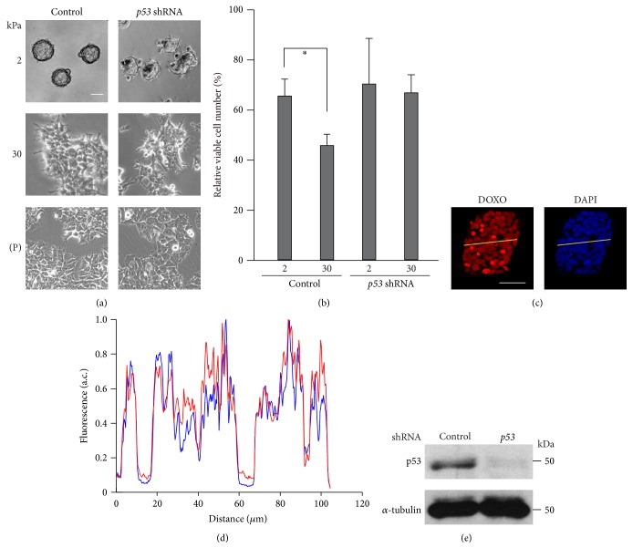
Rigid substrates enhance the inhibitory effect of doxorubicin on cell growth in a p53-dependent manner. (a-b) MCF-7 cells infected with a control or p53 shRNA-expressing retrovirus were cultured on substrates with elasticities of 2 and 30 kPa or on plates (P; ~106 kPa). (a) Phase contrast images of the cells were obtained with an inverted microscope (Olympus CKX41). Scale bar, 50 μm. (b) The cells were treated with doxorubicin (DOXO; 1 μg/mL) for 24 h, and the number of viable cells was counted using trypan blue exclusion-based cell staining. In each condition, the number of doxorubicin-treated cells was normalized to that of nontreated cells. Each bar represents the mean ± standard deviation (SD); n = 4. Asterisks, p < 0.05. (c-d) Accumulation of doxorubicin into cells cultured on a substrate with an elasticity of 2 kPa was evaluated. (c) Doxorubicin incorporated into cells was visualized using its autofluorescence. Z-stack images with an interval of 1.0 μm were obtained using a confocal microscope. Projected images for doxorubicin (red) and nuclei (DAPI) are shown. Scale bar, 50 μm. (d) The fluorescence intensity of the yellow line drawn across the spheroid was plotted. Intensity values were normalized with respect to the maximum value in each profile. (e) Extracts from control and p53 shRNA-expressing cells cultured on plates were subjected to immunoblot analysis with antibodies against p53 and α-tubulin as a loading control.

