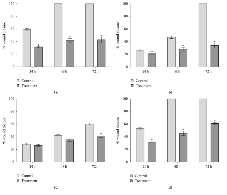
Figure 8.
Cell migration analysis using a wound-healing assay. HeLa (a), SiHa (b), CaSki (c), and C33A (d) cells were tested in 6-well plates (2.5 × 10⁴ cells/well) after scratching in the absence (negative control) and presence of apigenin. The results were calculated by comparing wound closure after 24, 48, and 72 h with the measurements taken at the initial time, and data are shown as the mean ± SD of three independent experiments conducted in triplicate. ∗P < 0.05 versus the negative control.

