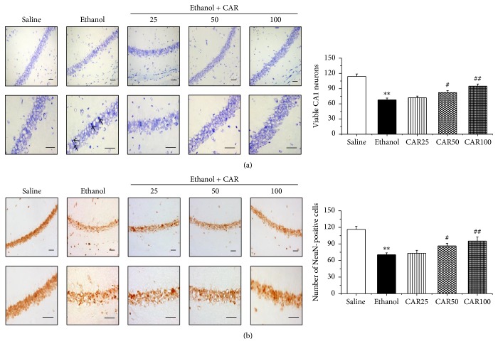
Figure 4.
Nissl staining and NeuN immunohistochemistry showed the protective effects of carvacrol on ethanol-induced neuronal impairment of the hippocampal CA1 region in C57BL/6 mice. (a) Representative photomicrographs of Nissl staining for surviving neurons in hippocampal CA1 region, and the statistical analysis of the surviving cells in each group. Arrows indicated the shrunk dark damaged neurons. (b) Representative immunohistochemical photomicrographs of NeuN in mouse hippocampus, and the statistical analysis of the NeuN-immunopositive cells in each group. There were fewer NeuN and Nissl-positive neurons in the ethanol group than in the saline group. With treatment with different doses of carvacrol, NeuN and Nissl-positive neurons were abundant in the CA1 region compared with the ethanol group. Up panel is lower magnification image (200x) and down panel is higher magnification image (400x) of CA1 pyramidal neurons. Scale bar: 50 μm. The data are expressed as the mean ± SEM (n = 6 per group). ∗∗P < 0.01 compared to saline group; #P < 0.05 and ##P < 0.01 compared to ethanol group.

