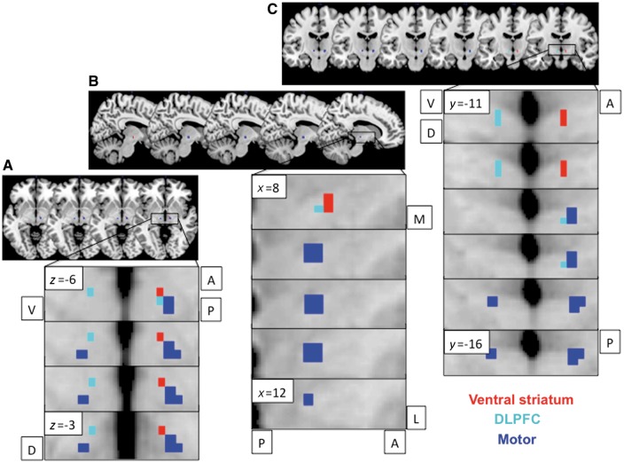
Figure 3.
Limbic-associative and motor connectivity dissociating anterior and posterior STN. Resting state functional connectivity of ventral striatal (red) and dorsolateral prefrontal cortex (DLPFC; cyan) seeds to anterior STN and primary motor cortex (blue) seed to posterior STN shown for axial (A), sagittal right (B) and coronal (C) STN slices. Bilateral ventral striatal seeds showed lateralized functional connectivity to right STN. The ventral striatal and dlPFC activations are shown at FWE P < 0.05 and motor activations are shown at FWE P < 0.001 with an STN mask on a standard MNI template. The different thresholds were used for illustration purposes as motor activity at lower threshold otherwise activated the entire STN. A = anterior; P = posterior; V = ventral; D = dorsal.

