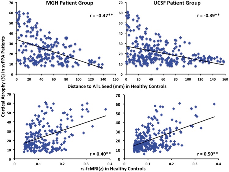
Figure 5.
Strength of connectivity of distributed cortical regions with the temporal pole in healthy adults predicts magnitude of atrophy within distributed cortical regions in svPPA patients. Within each region of interest the Euclidean distance to the temporal pole (top), and the strength of its functional connectivity with the temporal pole in young healthy adults (bottom) predicted the magnitude of cortical atrophy in both svPPA patient cohorts (**P < 0.001). The region of interest containing the temporal pole seed for the rs-fcMRI map was excluded from all correlation analyses.

