
Figure 2.
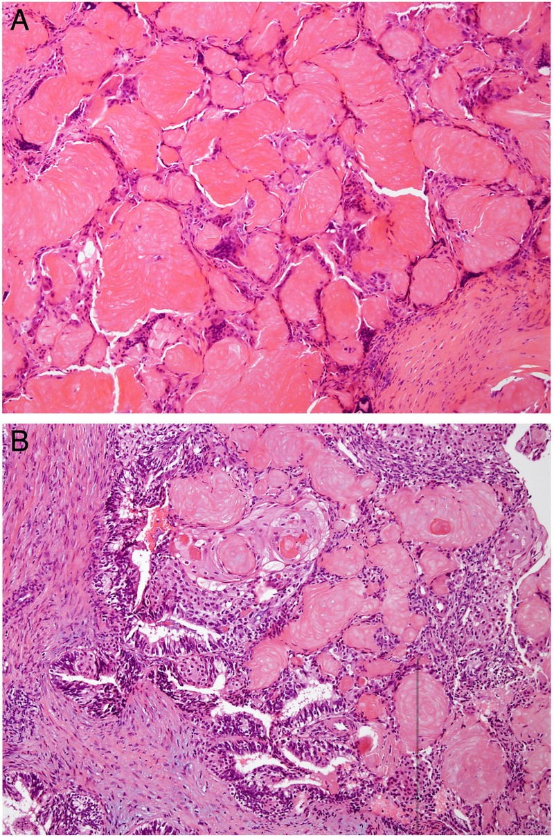
(A) H&E-stained section of the postradiotherapy vaginal lesion with nests of eosinophilic ‘ghost cells’ showing outlines of necrotic cells without retained nuclei surrounded by foreign body type giant cells mimicking a pilomatrixoma. (B) H&E-stained section of the first vaginal recurrence showing a central focus of endometrioid adenocarcinoma with squamous metaplasia and adjacent foci of acellular ghosts of squamous epithelium identical to those in the second lesion.