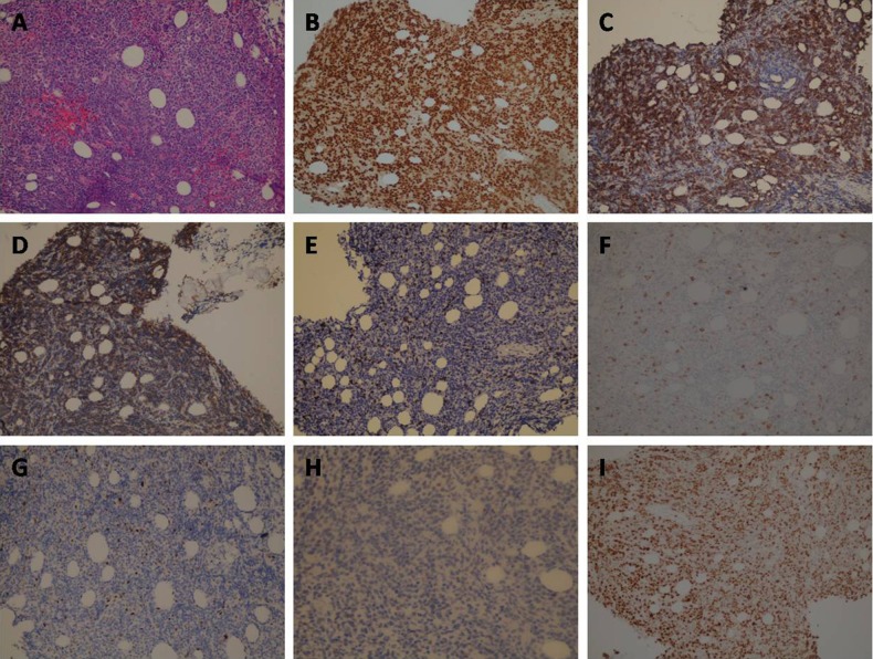
Figure 4.
(A) Proliferation of large cells with moderate cytoplasm, large vesicular nuclei and prominent nucleoli (×200); (B) positive staining for PAX-5 (×200); (C) positive staining for BCL-2 (×200); (D) positive staining for CD-20 (×200); (E) negative staining for CD-3 (×200); (F) negative staining for CD-5 (×200); (G) negative staining for BCL-6 (×200); (H) negative staining for cyclin-D1(×200); (I) Ki67 immunostaining showed a proliferation index of ∼70% (×200).

