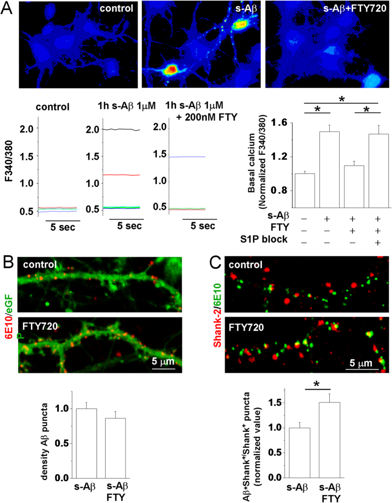
Figure 1. FTY720 protects neurons from s-Aβ-induced calcium dysregulation by altering the binding of s-Aβ to neurons.
Basal [Ca2+]i was measured in 9DIV neurons, loaded with the ratiometric calcium dye Fura-2 and expressed as F340/380 fluorescence. (A) Representative pseudocolor images of basal [Ca2+]i in control neurons and in neurons exposed to s-Aβ alone or in combination with FTY720 for 1 h. Representative traces and quantification of basal [Ca2+]i are shown below. Values are normalized to control (the Kruskal–Wallis ANOVA, P = 0.002; Dunn’s test for comparison among groups, P = 0.05; N = 4). (B) Representative confocal images of 14DIV eGFP+ mouse neurons exposed to s-Aβ alone or in combination with FTY720, fixed and stained with the anti-Aβ antibody 6E10. Quantification of Aβ binding to neurons is shown below. The histogram shows density of Aβ puncta bound to neurons, (number of Aβ/eGFP colocalizing puncta normalized over eGFP area) (Mann-Whitney Rank Sum Test, P = 0.153; N = 3). (C) Representative confocal images of mouse neurons treated as in (B) and probed for 6E10 and the postsynaptic density marker shank-2. The panel below shows the fraction of Shank-2 puncta with bound Aβ (Mann-Whitney Rank Sum Test, P = 0.043; N = 3).

