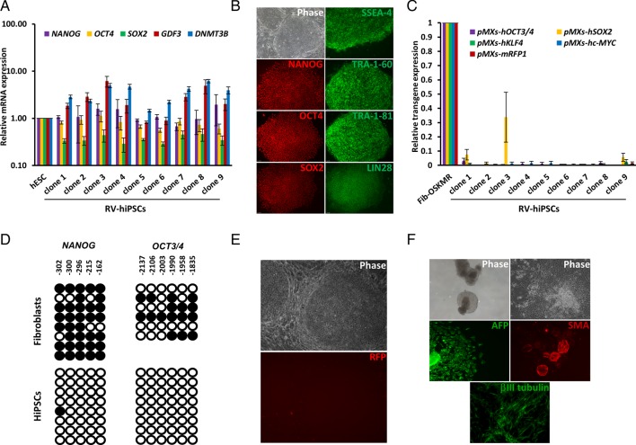
Fig. 3.
Characterisation of RV-hiPSC clones isolated based RV-Tg silencing and hESC-like morphology. (A) Real time PCR analysis of pluripotency markers in the isolated RV-hiPSC clones. The fold-change was calculated relative to the expression levels in hESCs (n=2). (B) Immunofluorescence analysis of pluripotency markers in the isolated clones. (C) Real-time PCR analysis of expression of retroviral transgenes in the clones (n=2). The fold-changes were calculated relative to the expression levels in fibroblast transduced with OSKMR (Fib-OSKMR). (D) Bisulfite sequencing results of OCT4 and NANOG promoters in fibroblasts and established RV-hiPSC clones showing hypomethylation of these regions. (E) Microscopic image of an established RV-hiPSC clone confirming the stable silencing of transgenes throughout the culture. (F) In vitro differentiation of established hiPSC clones. Data from a representative clone is shown. hiPSCs formed cystic embryoid bodies (EBs) in suspension culture and these EBs were differentiated further in an adherent culture into the cells expressing markers characteristic of three germ layers – endoderm (α-feto protein, AFP), mesoderm (α-smooth muscle actin, SMA) and ectoderm (βIII-Tubulin). All images are at 10× magnification. Data represented as mean±s.d.

