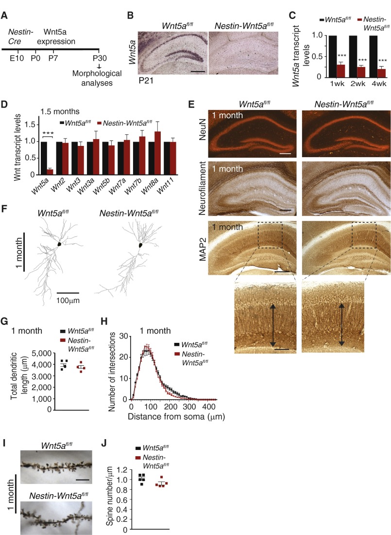
Fig. S1.
Wnt5a is dispensable for hippocampal development. (A) Schematic of the strategy for assessing effects of embryonic Wnt5a deletion on neuronal morphology, using Nestin-Cre transgentic mice. (B) Wnt5a transcript is undetectable in the P21 Nestin-Wnt5afl/fl hippocampus. (Scale bar, 500 μm.) (C and D) qPCR analyses shows substantial depletion of Wnt5a mRNA in Nestin-Wnt5afl/fl hippocampus, whereas other Wnts are unaltered. Results are means ± SEM from n = 7 mice per genotype for 1 and 2 wk, and n = 8 mice per genotype for 4 wk, and from six independent experiments for D. ***P < 0.001, two-tailed t test. (E) Hippocampal cyto-architecture, axonal projections, and CA1 dendritic layers are unaffected in Nestin-Wnt5afl/fl mice at 1 mo, revealed by immunostaining with antibodies against NeuN, neurofilament, and MAP2. Lower panels indicate higher magnification images of the boxed regions. [Scale bars, 400 μm (Upper) and 200 μm (Lower).] (F) Golgi staining and dendritic reconstructions show comparable arbor sizes in CA1 pyramidal Nestin-Wnt5afl/fl and control neurons at 1 mo. (Scale bar, 100 μm.) (G and H) Quantification of dendritic lengths and dendrite complexity, using Sholl analysis, in 1-mo-old Nestin-Wnt5afl/fl mice and Wnt5afl/fl littermate controls. Results are mean ± SEM from five neurons traced per animal and a total of four mice per genotype, two-tailed t test. (I and J) Spine densities in CA1 apical dendrites are normal in Nestin-Wnt5afl/fl mice at 1 mo. (Scale bar, 5 μm.) Results are mean ± SEM from five mice per genotype.

