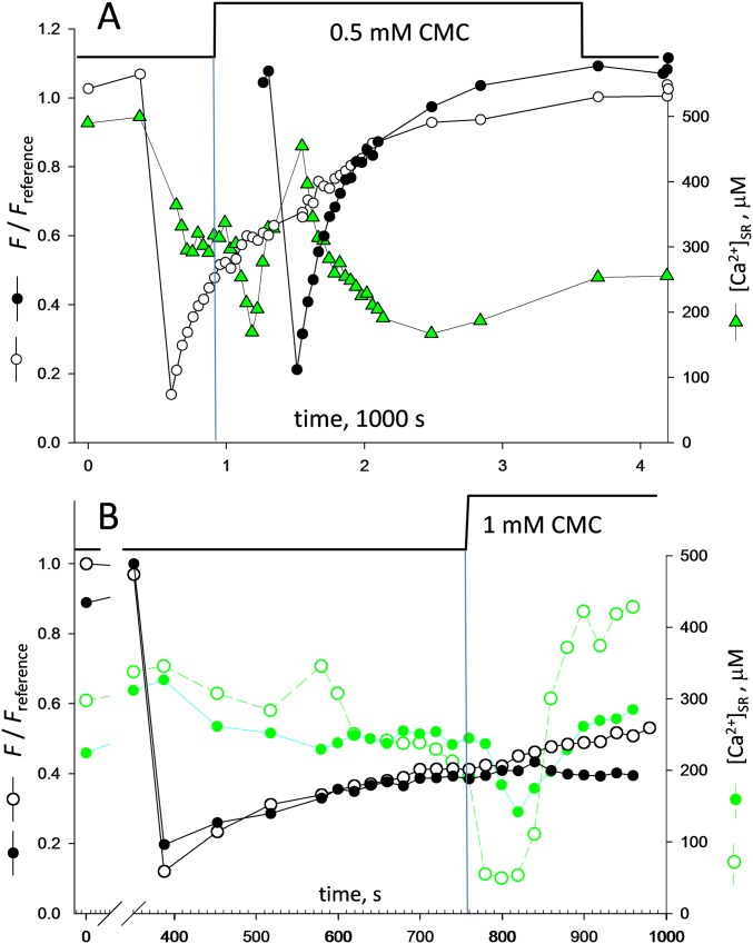
Fig. S5.
Examples of oscillations in [Ca2+]SR (supports Fig. 3D). (A) Example of oscillation of moderate amplitude in [Ca2+]SR. The oscillation was observed after the decay in [Ca2+]SR caused by the application of a depleting solution with a moderate concentration of 4-CMC. Compared with the case illustrated in Fig. 3D, the initial dip in [Ca2+]SR is less steep, and so is the ensuing surge. (B) Two myofibers were imaged in the same field and were bleached simultaneously. Upon application of a depleting solution, the fibers responded differently, perhaps because the depleting drug reached them at different rates. In both fibers, [Ca2+]SR oscillated (green symbols, open or filled); again, the depth and steepness of the initial decay correlated positively with the reach of the surge that followed. Note also that FRAP recovery, which had progressed to a similar extent in both fibers before depletion, evolved differently afterward, with fluorescence undergoing further recovery only in the fiber that experienced the greater oscillation (open circles).

