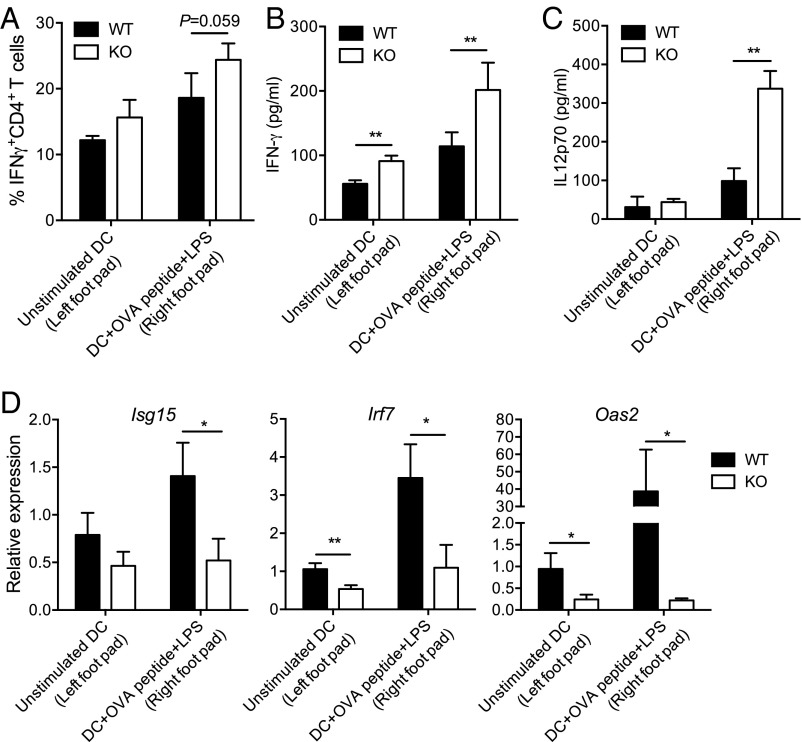
Fig. 5.
In vivo function of DCIR in Th1 immunity. OT-II T-cell receptor transgenic T cells were purified and injected into C57BL/6J recipients. Two days later, mice were challenged in the footpads with OVA peptide-pulsed or unpulsed WT or DCIR-deficient DCs. After 5 d of priming, popliteal LN cells were harvested from recipients. (A) Percentage of IFNγ-producing CD4+ OVA-specific (OT-II) Th1 cells adoptively transferred into C57BL/6J recipients and primed in vivo with peptide-pulsed or unpulsed DCs. (B and C) ELISA quantification of IFNγ (B) and IL-12–p70 (C) in LN lysates from recipient mice. (D) RT-qPCR quantification of gene expression of the ISGs Isg15, Irf7, and Oas2 in LN lysates from recipient mice. Data are presented as means ± SEM of five biological replicates. Statistical analysis was performed using Student’s t test. *P < 0.05; **P < 0.01.

