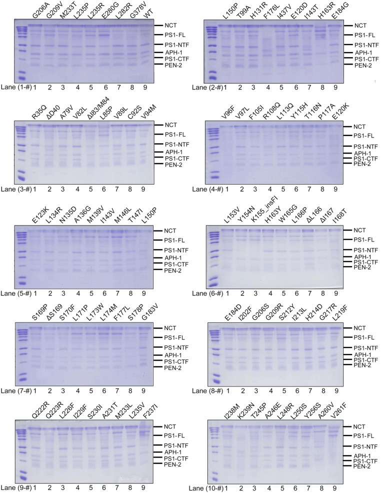
Fig. S2.
WT γ-secretase and variants used in this study. WT γ-secretase and variants are visualized on SDS/PAGE gels by Coomassie staining. For most variants, five polypeptide bands are clearly visible, representing Nicastrin (NCT), the N-terminal fragment (NTF) of PS1, APH-1aL, the C-terminal fragment (CTF) of PS1, and PEN-2. Each variant is identified by a specific PS1 mutation labeled at the top of the lane. Each lane is identified by two numbers, with the first representing the gel and the second specifying location within the gel. For example, WT γ-secretase is in lane 1-9 (gel 1, lane number 9).

