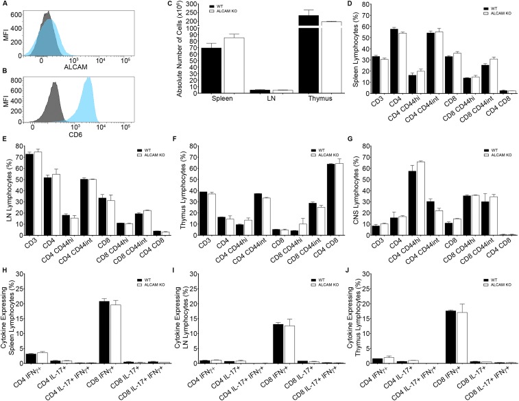
Fig. S1.
Expression of (A) ALCAM and (B) CD6 on resting memory CD4+ T lymphocytes isolated from naïve WT splenocytes, as assessed by flow cytometry. Mean fluorescence intensity of ALCAM (blue) or CD6 (blue) and their respective isotype control (gray). (C) Absolute number of leukocytes in the spleen, LN, and thymus of naïve ALCAM KO mice and their WT littermates. Percentage of lymphocytes isolated from the spleen (D), the LNs (E), the thymus (F), and the CNS (G) of naïve ALCAM KO and WT mice positive for the extracellular marker CD3, CD4, CD8, and CD44, as assessed by flow cytometry. Percentages of CD4+ and CD8+ cells are gated on CD3+ T lymphocytes. Percentages of CD44+ cells are gated on CD4+ or CD8+ T lymphocytes. Data shown are the mean ± SEM of 3–9 animals per time point. Percentage of CD4+ or CD8+ T lymphocytes isolated from the spleen (H), the LNs (I), and the thymus (J) of naïve ALCAM KO and WT mice expressing the cytokines IFNγ and/or IL-17, as assessed by flow cytometry. Data shown are the mean ± SEM of 3–9 animals per time point.

