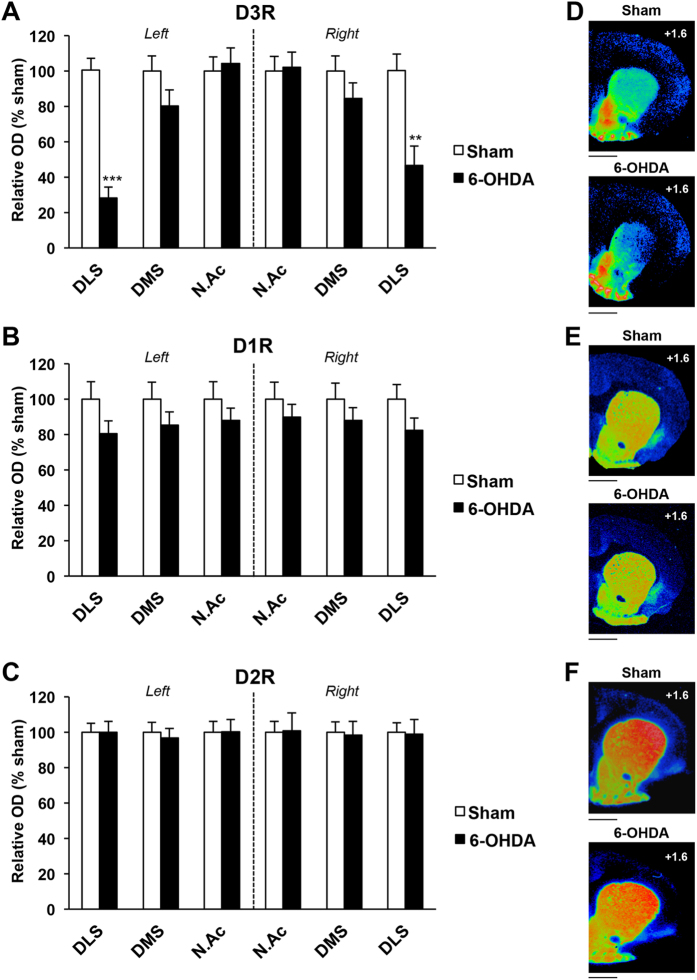
Figure 2. Bilateral 6-OHDA SNc lesion induces a selective decrease of D3R expression in dorsolateral striatum.
(A–C), Mean ± SEM optical density (expressed as arbitrary units) of D3R, D1R and D2R receptor binding density at striatal level, as measured by semi-quantitative autoradiography in sham and 6-OHDA lesioned rats. SNc lesions induced a marked decrease of D3R binding, specifically within the dorsolateral striatum. Two-way ANOVAs and post-hoc analyses with the method of contrasts were used. **p < 0.01, ***p < 0.001, sham (n = 8) vs 6-OHDA (n = 7). DLS: dorsolateral striatum; DMS: dorsomedial striatum; NAc: nucleus accumbens. (D–F) Photographs of autoradiograms obtained at striatal level for sham and 6-OHDA–lesioned rats, colorized using Autoradio V4.03 Software. D, D3R; E, D1R; F, D2R. AP levels are indicated from bregma. Scale: 1mm. AP: Anteroposterior.

