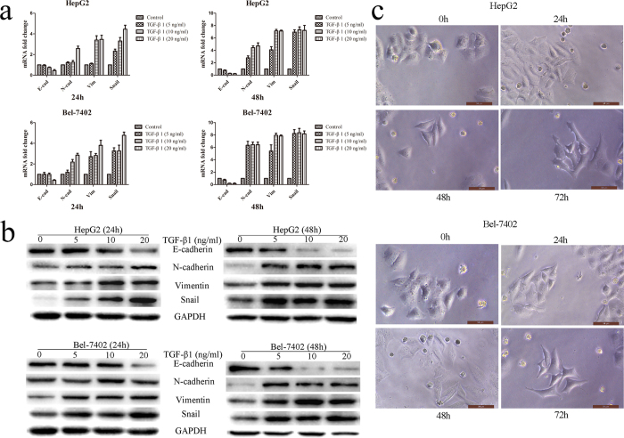
Figure 3. TGF-β1 induced EMT progress and restored migration, invasion abilities suppressed by Neferine.
(a,b) TGF-β1 induced EMT progress. HCC cells were treated with 5 ng/ml, 10 ng/ml, 20 ng/ml TGF-β1 for 24 hrs or 48 hrs to induce EMT. mRNA and protein expression of EMT biomarkers (E-cadherin, N-cadherin & Vimentin), and EMT promoting transcription factor (Snail) was determined by qRT-PCR and by Western blot. Original blots of high-contrast blots are presented in Supplementary Fig. S4. (c) Morphological changes of HCC cells treated with 10 ng/ml TGF-β1 for different times. TGF-β1 changed cells morphology from pebble-like epithelial to dispersed, spindle-like mesenchymal and pseudopodium stretching. (Original magnification: ×400).

