Figure 1.

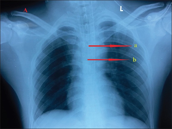
A chest radiography with polyvinyl chloride endotracheal tube in place. Arrow “a” is placed at the distal end of radiopaque line which will correspond to endotracheal tube tip. Arrow “b” is placed at carina.

A chest radiography with polyvinyl chloride endotracheal tube in place. Arrow “a” is placed at the distal end of radiopaque line which will correspond to endotracheal tube tip. Arrow “b” is placed at carina.