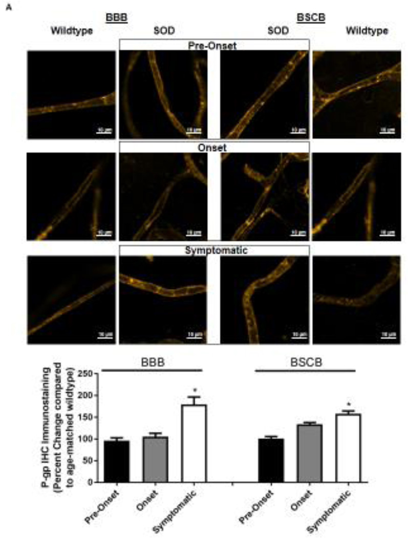
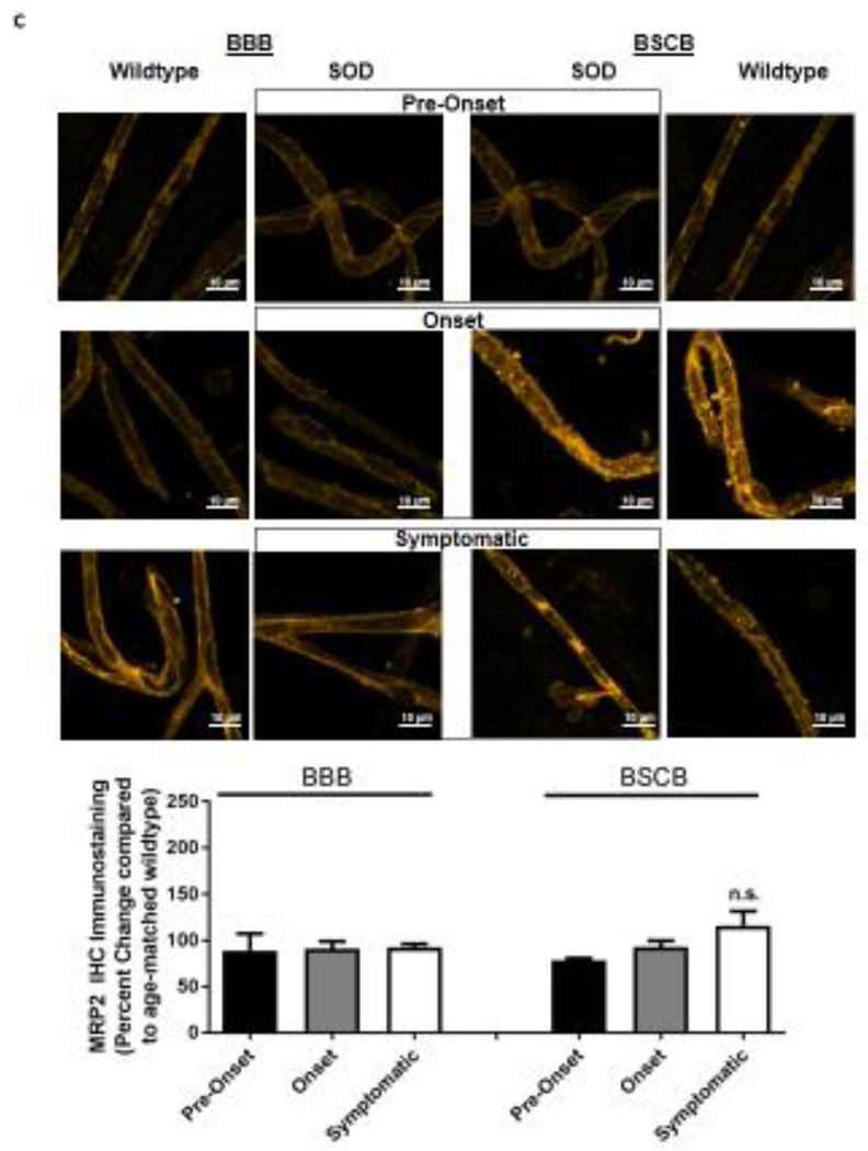
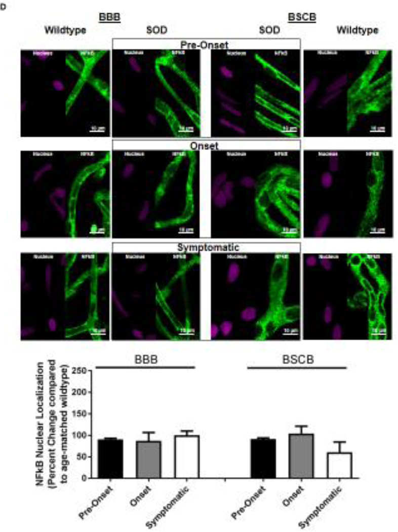
Figure 2. Immunohistochemical staining of P-gp, BCRP, Mrp 2 and NFkB in brain and spinal cord capillaries of SOD rats at ALS pre-onset, symptomatic onset and symptomatic stage and age-matched wildtype rats.
Representative confocal images and percent expression change of (A) P-gp, (B) BCRP, (C) MRP2, and D) NFkB in brain and spinal cord capillaries. Nuclear staining was achieved using DRAQ5 (left side of each representative image). For each protein, at least 10 capillaries per at least two capillary preparations (pooled tissue from at least four rats) were analyzed from each ALS stage of SOD rats and age-matched wildtype rats. Percent change (normalized to age-matched wildtypes) of protein immunohistochemical staining are presented with S.E.M.. Scale bars in confocal images indicates 10 µm. One-way ANOVA with Dunnett’s multiple comparisons post-hoc test was used. *Post-hoc test with p <0.05 was considered a significant difference compared to capillaries of SOD rats at pre-onset stage. n.s. = nonsignificant compared to capillaries exposed to pre-onset rats.


