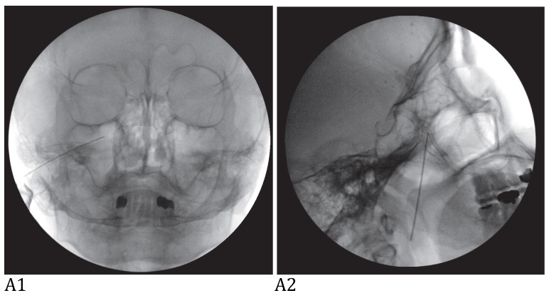
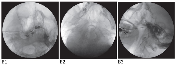
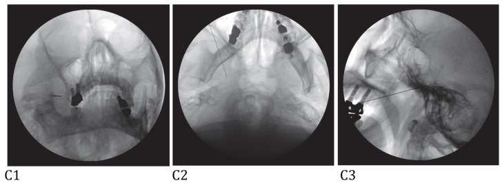
Figure 1.
A: Maxillary nerve block. A needle tip is located right lateral to foramen rotundum on the anteroposterior view (A1) and pterygopalatine fossa on the lateral view (A2). B: Mandibular nerve block. A needle tip is placed at the midportion of the foramen ovale on the anteroposterior oblique view (B1). At this point, a needle is advanced a few millimeters medially to elicit paresthesia in the V3 innervated area. When the proper paresthesia is achieved, a needle tip should be in a cross point of the lateral one-third of the perpendicular line and the midhorizontal line of the foramen ovale on the submentomandibular view (B2). On the lateral view, a needle is placed in the entrance of foramen ovale at the margin of petrous bone (B3). C: Combined maxillary and mandibular nerve block. A needle tip is placed at the midportion of the foramen ovale on the anteroposterior oblique view (C1). At this point, a needle is advanced a few millimeters cephalad passing foramen ovale on the submentomandibular view (C2). On the lateral view, a needle is placed passing foramen ovale a few millimeters under the clival line (C3).


