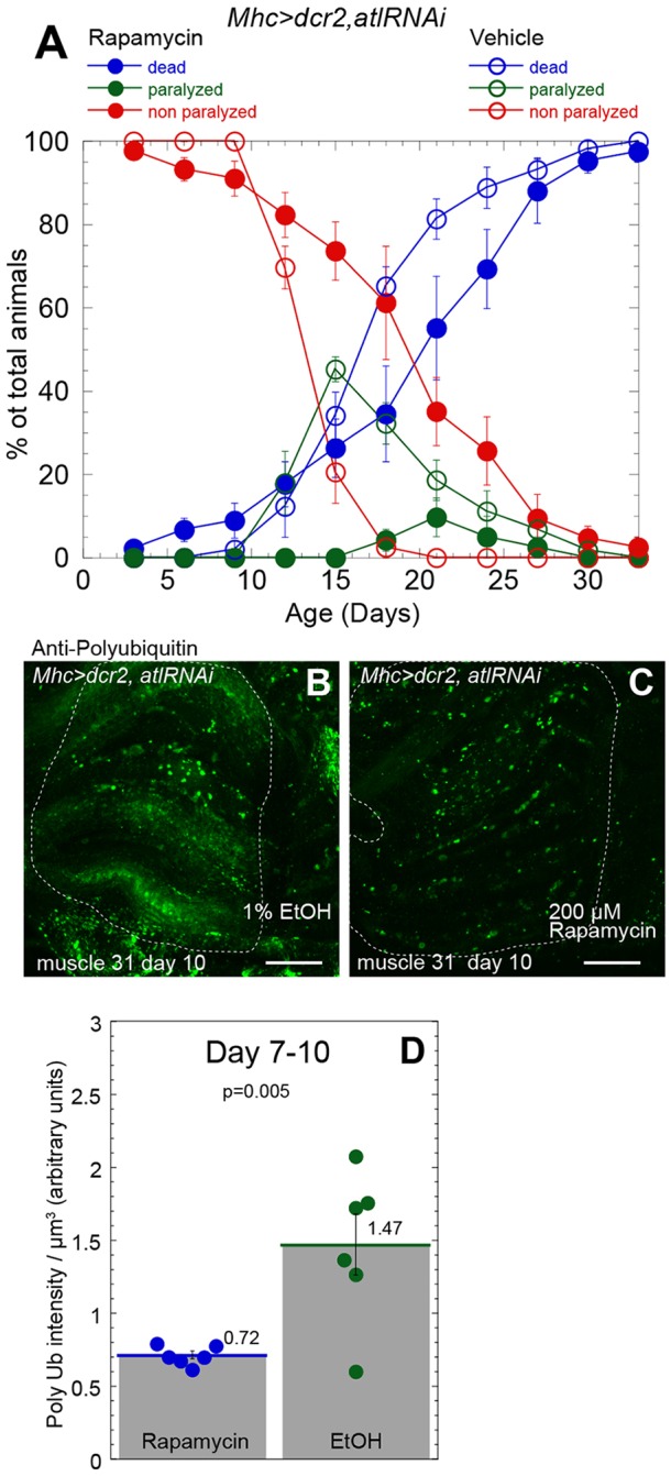
Fig. 8.

Rapamycin administration partially suppresses locomotor and viability deficits and polyubiquitin aggregate accumulation conferred by muscle atl knockdown. All animals analyzed were males that had been reared as mating pairs. (A) Mhc>dcr2,atlRNAi flies, maintained at 25°C as single mating pairs, were fed with food containing 200 µM rapamycin (solid circles) or 1% EtOH vehicle (open circles) immediately following eclosion. Viability (blue and red circles) and paralysis (green circles) were measured. The means±s.e.m. of the percentage of flies living, paralyzed or dead from five replicates (40 animals) of Mhc>Dcr2, atlRNAi flies that had been treated with rapamycin and five replicates (41 animals) of Mhc>Dcr2,atlRNAi flies that had been treated with vehicle are shown. (B,C) Dissected thoracic tissue from 10-day-old Mhc>dcr2,atlRNAi adults that had been fed immediately following eclosion with either vehicle (1% EtOH, B) or 200 µM rapamycin (C) was fixed and stained with an anti-ubiquitin antibody. Coxal muscle 31 is outlined. Scale bars: 25 µm. Maximum intensity z-projections from a confocal z-stack are shown. (D) Quantification of polyubiquitin (Poly Ub)-staining pixel intensities per µm3 in each muscle from flies fed with rapamycin (blue circles) or vehicle (green circles) following staining with an anti-ubiquitin antibody. Images from 7- and 10-day-old flies were pooled. Means±s.e.m. are above each bar. P=0.005 by Student's t-test.
