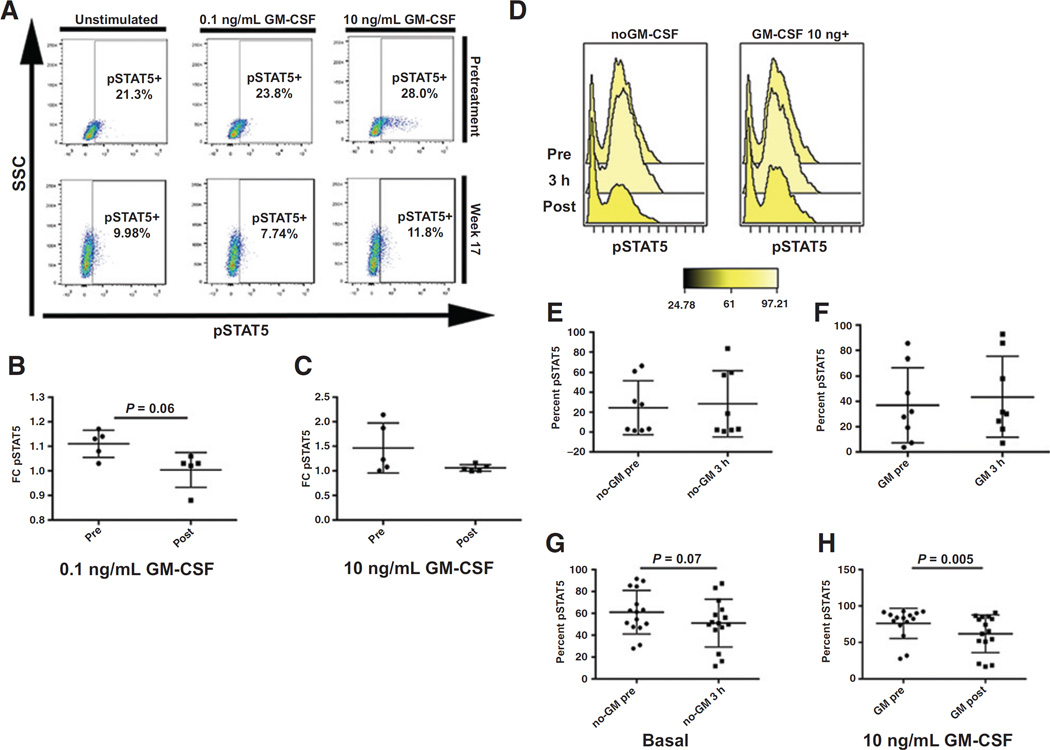
Figure 4.
Ruxolitinib suppresses GM-CSF–dependent pSTAT5 activation. A, representative sample demonstrating change in pSTAT5 activation while on ruxolitinib therapy. Percent of cells in pSTAT5 gate are noted. B, fold change of GM-CSF 0.1 ng/mL stimulated pSTAT5 levels relative to basal pSTAT5 in pretreatment and week 17 (± 2 weeks) bone marrow samples (n = 5). C, fold change of GM-CSF 10 ng/mL stimulated pSTAT5 levels relative to basal pSTAT5 in pretreatment and week 17 (±2 weeks) bone marrow samples (n = 5). D–H, PIA results demonstrating the percent of THP-1 cells in the pSTAT5 gate comparing representative sample of THP-1 cells treated with patient plasma (D), basal pretreatment or screening samples (pre) with samples taken 3 hours after the first dose (3 hours; n = 11; E, GM-CSF 10 ng/mL stimulated pretreatment or screening samples (pre) with samples taken 3 hours after the first dose (3 hours; n = 11; F, basal pretreatment or screening samples (pre) with samples taken while patient was on study for an average of 10 weeks (post). (n = 15; G). H, GM-stimulated 10 ng/mL pretreatment or screening samples (pre) with samples taken while patient was on study for an average of 10 weeks (post). (n = 15).

