Figure 1.

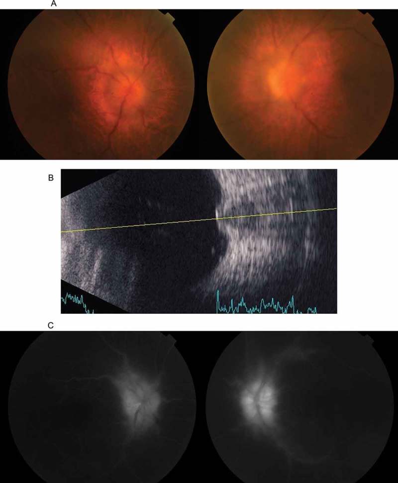
(A) A colour fundus photograph taken at the initial presentation, showing bilateral optic disc swelling. The haze is attributable to vitritis. (B) A B-mode ultrasonograph of the left eye, showing marked protrusion of the optic disc into the vitreous cavity. (C) Fluorescein angiography showing leakage from the optic disc.
