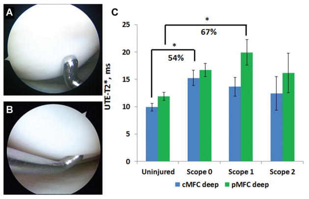
Figure 2.
Ultrashort echo time (UTE)–T2* in deep articular cartilage varies with the degree of arthroscopic softening among participants with intact articular surfaces (Kruskal-Wallis test, P = .01). (A) Arthroscopic image of the medial femoral condyle (MFC) of an anterior cruciate ligament (ACL)–reconstructed patient with firm and intact cartilage (Outerbridge grade 0). (B) The MFC of an ACL-reconstructed patient with intact and softened cartilage (Outerbridge grade 1). (C) ACL-reconstructed patients with intact and firm cartilage (grade 0) demonstrate significantly higher UTE-T2* values in deep central MFC cartilage (P = .01) compared with uninjured controls. In the posterior MFC, deep cartilage of ACL-reconstructed patients with softened but intact cartilage (grade 1) is significantly elevated compared with that of uninjured controls (P = .01). Error bars indicate standard error of the mean.

