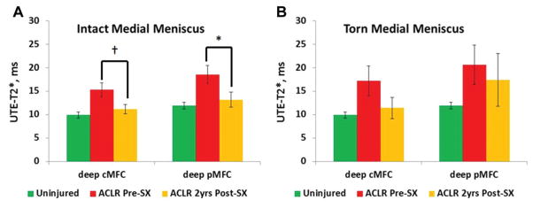
Figure 4.
Change in ultrashort echo time (UTE)–T2* values of articular cartilage at 2 years after anterior cruciate ligament reconstruction (ACLR) varied based on meniscus status at the time of surgery. (A) Mean UTE-T2* values decreased 29% for deep posterior medial femoral condyle (pMFC) cartilage (P = .01) and showed a trend toward a decrease for deep central MFC (cMFC) cartilage (P = .07) 2 years after ACLR in joints with intact medial menisci (n = 11). (B) In ACL-reconstructed joints with torn medial menisci, no significant change in the mean UTE-T2* value of the MFC was detected (n = 5; P = .14 and .5 for deep cMFC and deep pMFC, respectively). Error bars indicate standard error of the mean. *P <.05. †Denotes a trend.

