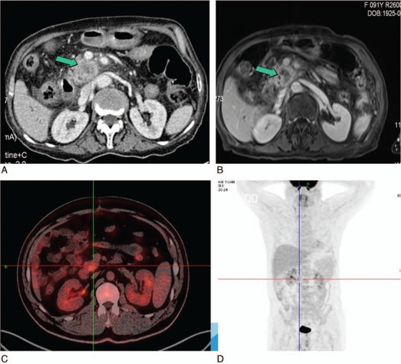
Figure 1.

(A) Abdominal contrast-enhanced CT and (B) contrast-enhanced MRI showed a 3 × 3 cm solid hypovascular tumor in the head of the pancreas (green arrow). (C, D) PET-CT showed a mass with SUVmax 11.6 located in the pancreatic head and no distant organ metastasis. CT = computed tomography, MRI = magnetic imaging resonance, PET = positron emission tomography.
