Figure 5.

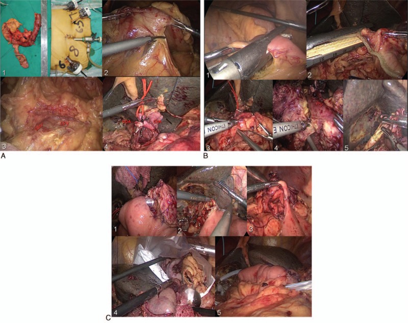
(A) 1 – Showing 5 trocars in a V-shape; 2, 3 – dividing the gastrocolic ligament, exposing the pancreas, SMV, and SPV; and 4 – ligating the GDA. (B) 1, 2 – The jejunum and stomach dissection (linear stapler); 3 – pancreatic neck division; 4 – dissecting the uncinate process of pancreas; and 5 – the bile duct division. (C) 1 – The end-to-side pancreaticojejunostomy (duct-to-mucosa); 2 – the end-to-side choledochojejunostomy; 3 – the gastrojejunostomy; 4 – the specimen removed; and 5 – placing 2 drainage tubes. GDA = gastroduodenal artery, SMV = superior mesenteric vein, SPV = splenetic vein.
