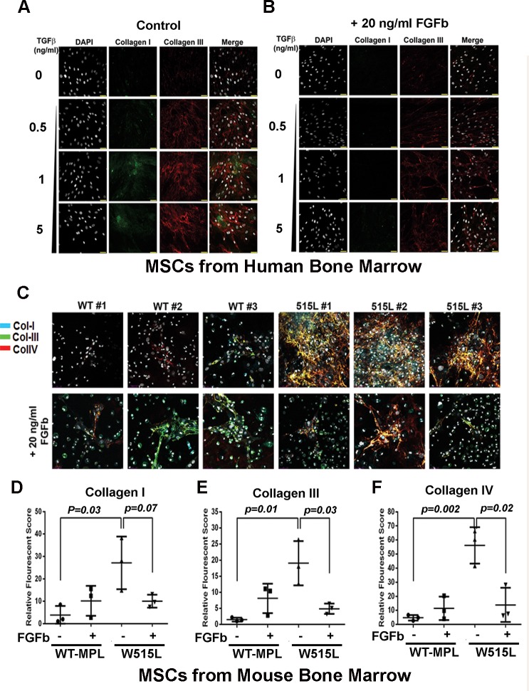
Fig 6. FGFb reverses the fibrotic phenotype of MPLW515L- MSCs.
FGFb and LIF maintain MSCs in a pluripotent state, but stimulate proliferation and expansion. (A, B) MSCs derived from human bone marrow were exposed to increasing concentrations (0.5, 1, 5 ng/ml) of TGFβ to enhance collagen I (green) and collagen III (red) in three-dimensional culture conditions in the absence (A) and presence of 20 ng/ml FGFb (B). (C) MSCs from three different mice expressing MPLWT or MPLW515L which has increased expression of collagen I, III, and IV. Presence of 20 ng/ml FGFb reduces collagen deposition in the culture after 72 hours. Cell numbers were increased after continuous culture in this growth factor (data not shown). Quantification of collagen I (D) collagen III, (E) collagen IV (F) fibers under each condition shown with p-values indicated based on analysis using a 2-sided t-test.

