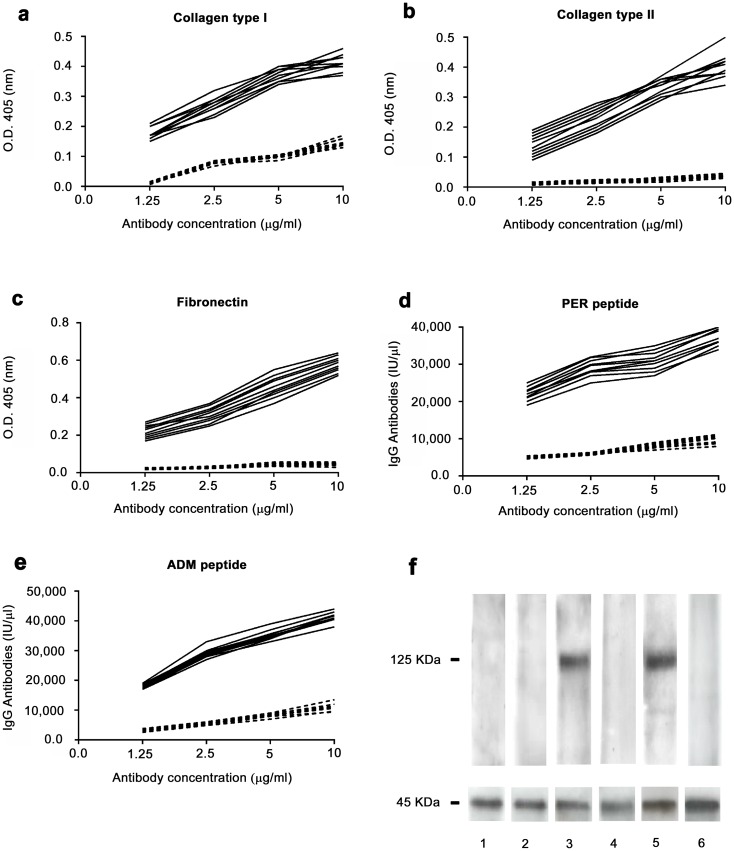
Fig 2. Binding of anti-peptide antibodies to autoantigens.
Binding of affinity purified anti-AS peptide antibodies to collagen type I (A), collagen type II (B), fibronectin (C), PER peptide (D), ADM peptide (E). Black continous line: antibodies affinity purified against the AS peptide from the sera of patients affected by AS. Dotted line: antibodies affinity purified against an irrelevant control peptide. A, B and C: ELISA assay. D and E: DELFIA assay. X axis: increasing antibody concentration by two fold ranging from 1.25 microgram/ml to 10 microgram/ml. Y axis: Optical Density values obtained at 405 nm wavelength for ELISA assay and IgG international units for DELFIA assay. F: western blot analysis of the binding of anti-peptide antibodies to ASAP1(molecular weight 125 KDa). Lane 1 and 2, antibodies affinity purified against an irrelevant control peptide probed with cell lysate from cells transfected with ASAP1 (lane 1) or from untransfected cells (lane 2); lane 3 and 4, antibodies affinity purified against the AS peptide probed with cell lysate from cells transfected with ASAP1 (lane 3) or from untransfected cells (lane 4); lanes 5 and 6, commercially available monoclonal antibody directed against ASAP1, probed with cell lysate from cells transfected with ASAP1 (lane 5) or from untransfected cells (lane 6). Lower panel of Fig 2F: immunoblot with actin demonstrates equal protein loading.

