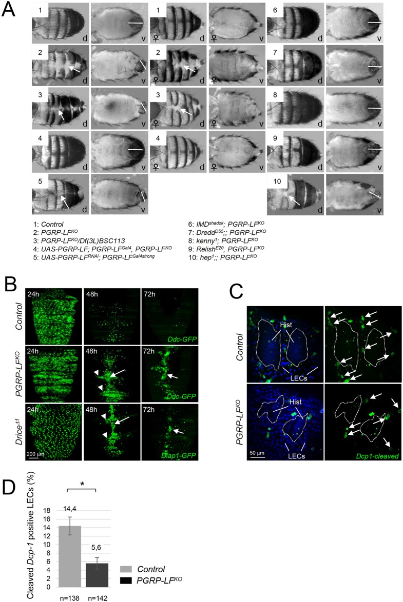
Fig 5. Adult PGRP-LF mutants display abdominal cuticle and male genitalia defects.
(A) Dorsal (d) and ventral (v) views of male or female abdomens showing incomplete epidermis differentiation (white arrow) and abnormal male genitalia orientation (white bar). (B) Elimination of larval epidermal (LECs) cells is not taking place in PGRP-LF mutants. LECs are labeled with the DdcGal4 driver combined with UAS-GFP. LECs are prominently distributed along the surface of the dorsal abdomen of control pupae at 24 hours APF, but by 48 hours APF most of LECs have been eliminated. By 72 hours APF, only very few intact LECs remains. At 48h and 72h APF, PGRP-LFKO and DriceΔ1 mutant pupae present many persistent LECs that accumulate along the midline of the abdomen (white arrow), and at segmental borders (arrowhead). (C) Defective LEC cell death in PGRP-LF mutant cuticle. Pictures show the lateral abdomen of 24h hours APF pupae stained with the anti-cleaved Dcp-1 antibody which labels caspase-activating LECs (arrows). The dashed lines indicate the boundary between the histoblasts and the LECs. (D) Quantification of dying LECs for 6 pupae 24h hours APF is shown in the histogram. For (D) histograms correspond to the mean value ± SD of six samples. Values indicated by symbols (*) are statistically significant (t-test, p < 0.05).

