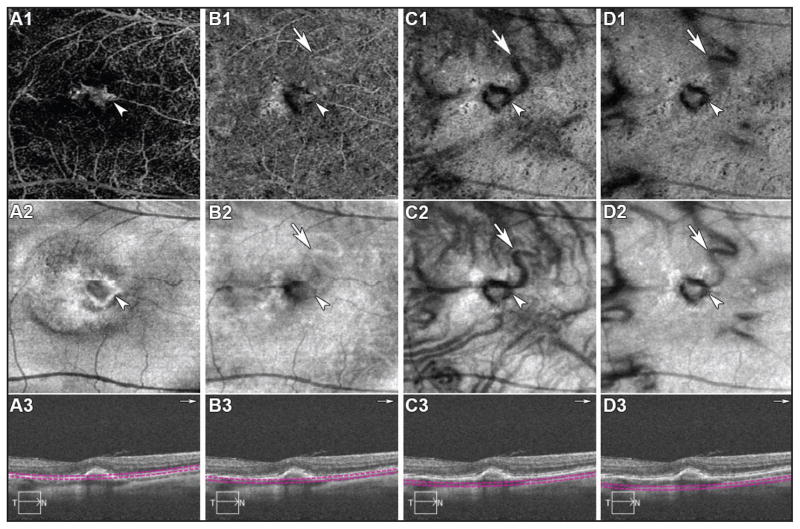
Figure 2.
Spectral-domain optical coherence tomography angiography (SD-OCTA) from the right eye demonstrates multiples depths of scans (A, B, C, D). OCTA (upper row), en face structural OCT (middle row), and cross-sectional OCT (lower row) are shown. (A, B, C, D) White head arrows indicate the choroidal neovascularization and white arrows indicate the feeder vessel in all scans.

