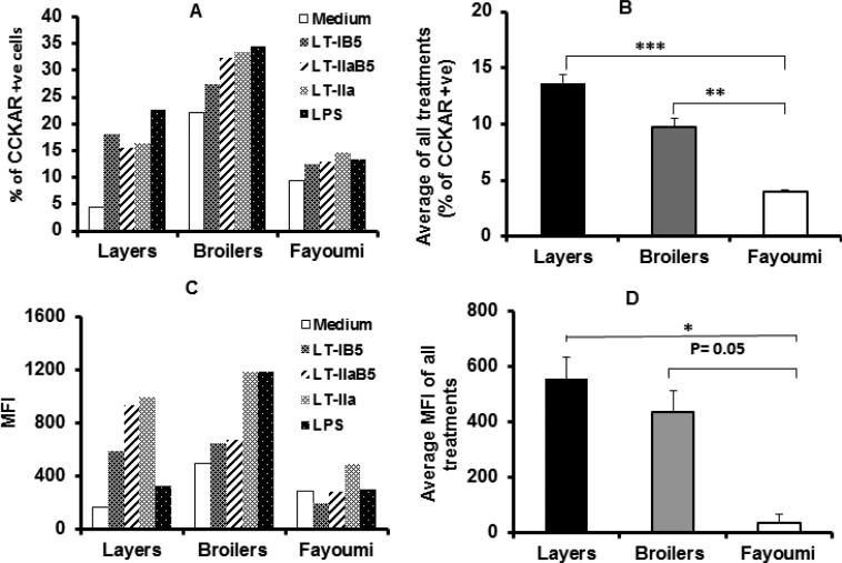
Figure 1.
Stimulation of chicken monocytes significantly increases CCKAR protein expression. PBMC were cultured with the E. coli enterotoxin LT-IIa or the B-subunit of LT-I and LT-II (LT-IB5 and LT-IIaB5, respectively), or LPS. Cells were harvested, and labelled with FITC anti-CCKAR and PE anti-KUL01 (for monocytes) and analyzed by flow cytometry. The percentage of cells expressing CCKAR was determined by gating out low FSC/high SSC cells (dead cells) [17] and platelets, and by gating on CCKAR+ and KUL01+cells. The total number of cells expression CCKAR (Figure 1A) and Mean Fluorescence Intensity (MFI) (Figure 1C) are shown. The results represent average of two experiments. In B and D, the average effects of all stimulants (− medium) on number of cells expressing CCKAR (Figure 1B) and level of CCKAR (Figure 1D) in the different breeds is compared. *, ** and *** denotes statistical significance (one-way Anova) with a p<0.05, p <0.01 and p<0.001, respectively.

