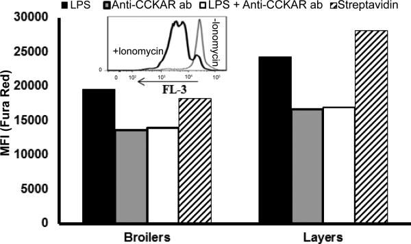
Figure 2.
Mobilization of Ca2+ following ligation of CCKAR. PBMC were isolated from pooled blood samples from 3 birds. To stimulate expression of CCKAR, cells were pre cultured for 24 h with LPS. Cells were then loaded with Fura Red Ca+2 dye. To induce mobilization of calcium, biotin conjugated anti CCKAR antibody and streptavidin were employed. Shown is a representative of one of two experiments with similar results. LPS indicates cells stimulated with LPS alone (no anti CCKAR); anti CCKAR indicates non LPS stimulated cells ligated with anti CCKAR + streptavidin; LPS + anti CCKAR ab indicates LPS stimulated cells ligated with anti CCKAR antibody + streptavidin, and streptavidin indicates cells to which streptavidin alone was added.

