. 2017 Jan 30;6:e19720. doi: 10.7554/eLife.19720

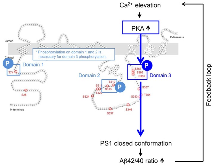
Figure 6. Mechanism of the Ca2+-triggered PS1 pathogenic conformational change.

The schematic image of the molecular events involved in the Ca2+-triggered pathogenic ‘closed’ conformational change and increase of the Aβ42/40 ratio. The elevated Ca2+ levels induce PKA activation, followed by the phosphorylation of PS1 at domain 1, domain 2 and domain 3. Domain 3 phosphorylation, particularly at S367, induces the PS1 pathogenic conformation that leads to increase in the Aβ42/40 ratio.

DOI: http://dx.doi.org/10.7554/eLife.19720.020

Figure 6.

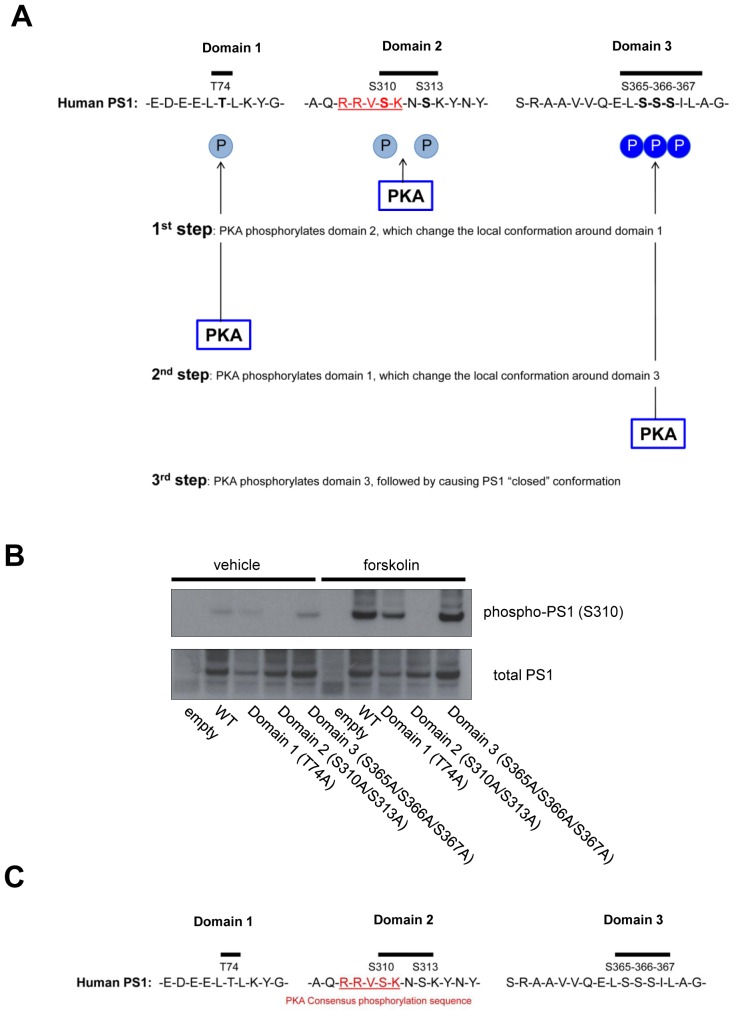
Figure 6—figure supplement 1. Model of the PKA-mediated PS1 phosphorylation.

Figure 6—figure supplement 1.

(A) Model. PKA first phosphorylates domain 2, followed by changing local conformation around domain 1. PKA then phosphorylates domain 1, which subsequently leads to local rearrangement around domain 3. Finally, PKA phosphorylates domain 3, causing PS1 pathogenic conformation. (B) The level of phosphorylated PS1 at S310 was compared between forskolin and vehicle-treated 7 W cells expressing WT, domain 1, 2 or 3 phospho-inhibited PS1 by Western blotting. (C) Amino acid sequences around domain 1, 2 and 3. Domain 2 includes the PKA substrate consensus sequence: R-R-V-S-K.