
Figure 1.
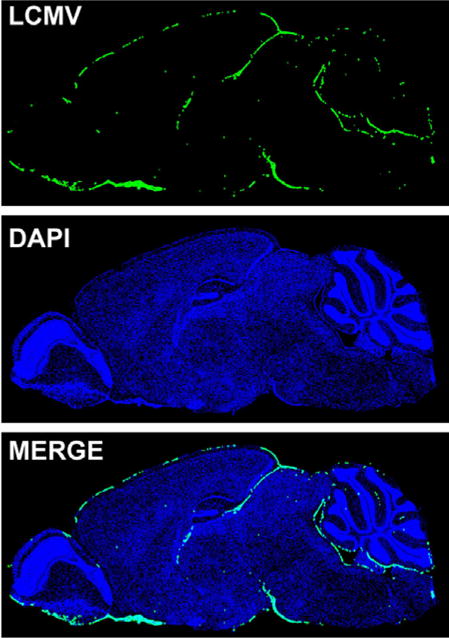
LCMV distribution during acute meningitis. The brain from an 8 week old C57BL/6 mouse infected with LCMV i.c. was harvested on day 6 post infection when severe seizures were apparent. Shown is the localization of LCMV (green) on a sagittal brain section. The virus was detected with a polyclonal anti-LCMV antibody and cell nuclei (blue) were stained with DAPI. Note that LCMV is distributed in the meninges with little to no infiltration into the brain parenchyma.