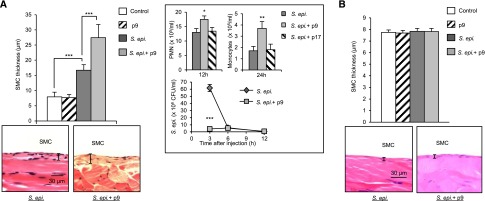
Figure 6.
The intensity of the TLR–mediated inflammatory episodes in addition to their number affects peritoneal fibrosis development. C57BL/6J mice (n=5 per group) were inoculated intraperitoneally (A) four or (B) two times at weekly intervals with (A) live or (B) heat–killed S. epidermidis (5×10⁸ CFU per mouse) or PBS (control) in the presence or absence of a TLR2-derived peptide (p9 or control p17; 20 μg per mouse). Four weeks after the last injection, histologic analysis of the peritoneal membrane was conducted as described in Figure 3. Representative fields (×40 magnification) are shown, and bar plots show the mean (±SEM) of the submesothelial compact zone (SMC) thickness for each group. (A, inset) At the indicated time points after the first injection, peritoneal lavages were obtained. PMN and monocyte numbers were determined by flow cytometry using anti-Ly6G– and anti-7/4–specific mAbs and bacterial titers by overnight culture on microbiologic agar plates. S. epidermidis and p9 versus S. epidermidis alone. *P<0.05; **P<0.01; ***P<0.01.

