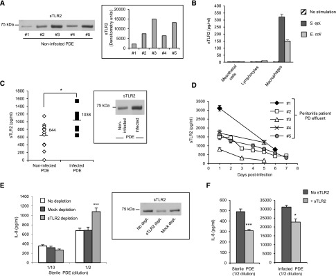
Figure 7.
sTLR2 is present in PDE from noninfected patients and can reduce PD–induced proinflammatory responses ex vivo. (A) Western blot analysis and densitometric scanning of sTLR2 detected in sterile PDE from different donors. (B) Levels of sTLR2 in the culture supernatants of peritoneal mesothelial cells, sterile PDE–isolated lymphocytes, and macrophages stimulated (16 hours) or not with heat–killed S. epidermidis (5×10⁸ CFU/ml) or E. coli (5×10⁷ CFU/ml). Results are from one experiment representative of three performed with cells from different donors. (C and D) Levels of sTLR2 in PDE from patients without or with ongoing peritoneal infection tested at (C) day 1 or (D) the indicated times postinfection. C, inset shows a representative sTLR2 Western blot profile in the PDE of one patient before and on day 1 of infection. In D, results are expressed as means (±SD) for each time point. *P<0.05. (E and F) Levels of IL-8 in the culture supernatant of (E) PBMC or (F) PDE leukocytes cultured overnight with the indicated dilutions of sterile or infected cellfree PDE (E) depleted of sTLR2, mock depleted, or not depleted or (F) supplemented with 500 ng/ml human recombinant sTLR2. Depletion in E was approximately 80% as estimated by Western blot. Results are from one experiment (±SD) representative of three performed with cellfree PDE from different donors. sTLR2 depleted versus mock depleted or and sTLR2 versus no sTLR2. *P<0.05; ***P<0.01.

