Figure 4.
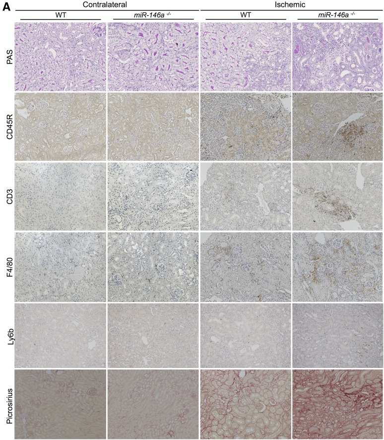
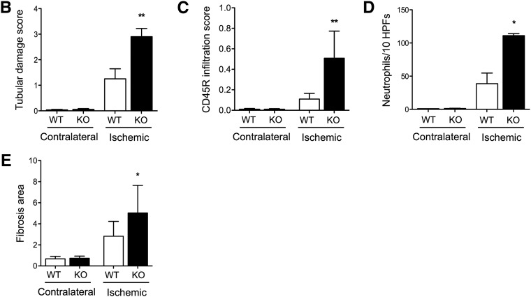
miR-146a deficiency increases tubular injury, interstitial inflammation, and fibrosis after renal IRI. (A) Representative sections of outer medullae from WT and miR-146a−/− mice at 14 days after reperfusion stained with periodic acid–Schiff (PAS) or immunostained for CD45R, CD3, F4/80, and Ly6b. Alternatively, collagen deposits were visualized by picrosirius red staining. Original magnification, ×200. (B) Semiquantitative analysis of tubular damage in injured kidneys of WT (n=6) and miR-146a−/− (n=6) mouse kidneys at day 14 post-IRI. (C) Semiquantitative analysis of CD45R+ leukocyte infiltration into the injured kidneys of WT and miR-146a−/− mice at day 14 post-IRI. (D) Quantification of infiltrating Ly6b+ neutrophils into the injured kidneys of WT and miR-146a−/− mice at day 14 post-IRI. (E) Quantification of WT and miR-146a−/− mouse kidneys stained with picrosirius red for visualization of collagen deposits. Data are shown as means±SEM; n=6–10 mice per group. Asterisks indicate comparisons of injured kidneys between WT and miR-146a−/− mouse. *P<0.05; **P<0.01.


