Figure 7.
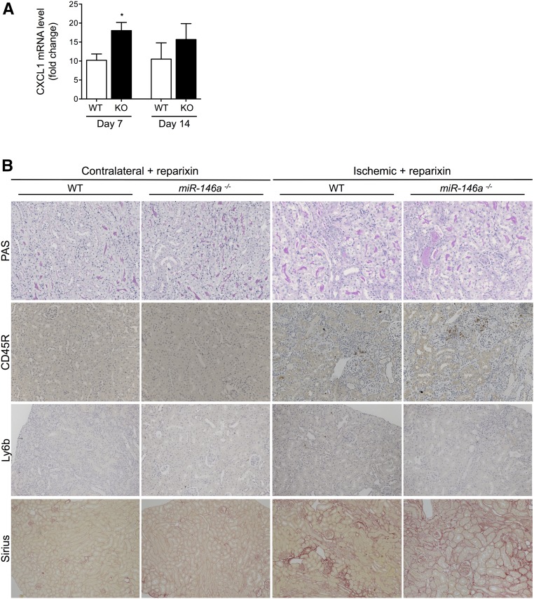
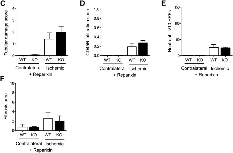
Reparixin prevents renal fibrosis in miR-146a−/− mice after IRI. (A) CXCL1 mRNA levels in WT and miR-146a−/− mouse kidneys 7 and 14 days post-IRI. Target mRNA levels were normalized to HPRT levels. (B) Representative sections of outer medullae from WT and miR-146a−/− mice at 14 days after reperfusion, with reparixin treatment from day 7 to day 14. Original magnification, ×200 for periodic acid–Schiff (PAS), picrosirius red staining, CD45R, and Ly6b immunostaining. (C) Semiquantitative analysis of tubular damage in reparixin–treated postischemic kidneys from WT (n=4) and miR-146a−/− mice (n=4). (D) Semiquantitative analysis of CD45R+ leukocyte infiltration into the injured kidneys of reparixin–treated WT and miR-146a−/− mice at day 14 post-IRI. (E) Quantification of infiltrating Ly6b+ neutrophils into the injured kidneys of reparixin–treated WT and miR-146a−/− mice at day 14 post-IRI. (F) Analysis of picrosirius staining in the renal interstitium of miR-146a−/− mice compared with WT mice. Data are shown as means±SEM; n=6 mice per group. Asterisk indicates comparisons of injured kidneys between WT and miR-146a−/− mice. *P<0.05.


