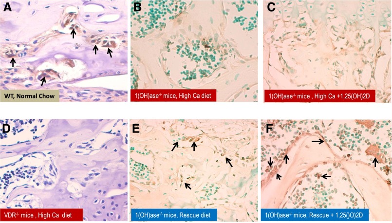
Figure 3.
Increased VDR expression in osteoblasts is induced by 1,25(OH)2D and further amplified by increasing serum calcium. Representative sections of VDR expression in bone analyzed by immunohistochemistry of femur from WT, VDR−/−, and 1(OH)ase−/− mice. The decalcified bone tissue was paraffin-embedded and stained for antibodies to VDR. Positive labeling for VDR in WT mice on normal chow is clearly evident in brown and denoted by black arrows (A). VDR expression in VDR−/− mice on a rescue diet was a negative control and showed no staining (D). VDR expression in femurs of 1(OH)ase−/− mice on a high-Ca diet contained undetectable to low levels of VDR (B), and no change was observed after treatment with exogenous 1,25(OH)2D (C). VDR expression in 1(OH)ase−/− mice was upregulated by a rescue diet (E) and increased further after exogenous treatment with 1,25(OH)2D (F). Original magnification, ×300 in A and D; ×200 in B, C, E, and F.

