Figure 6.
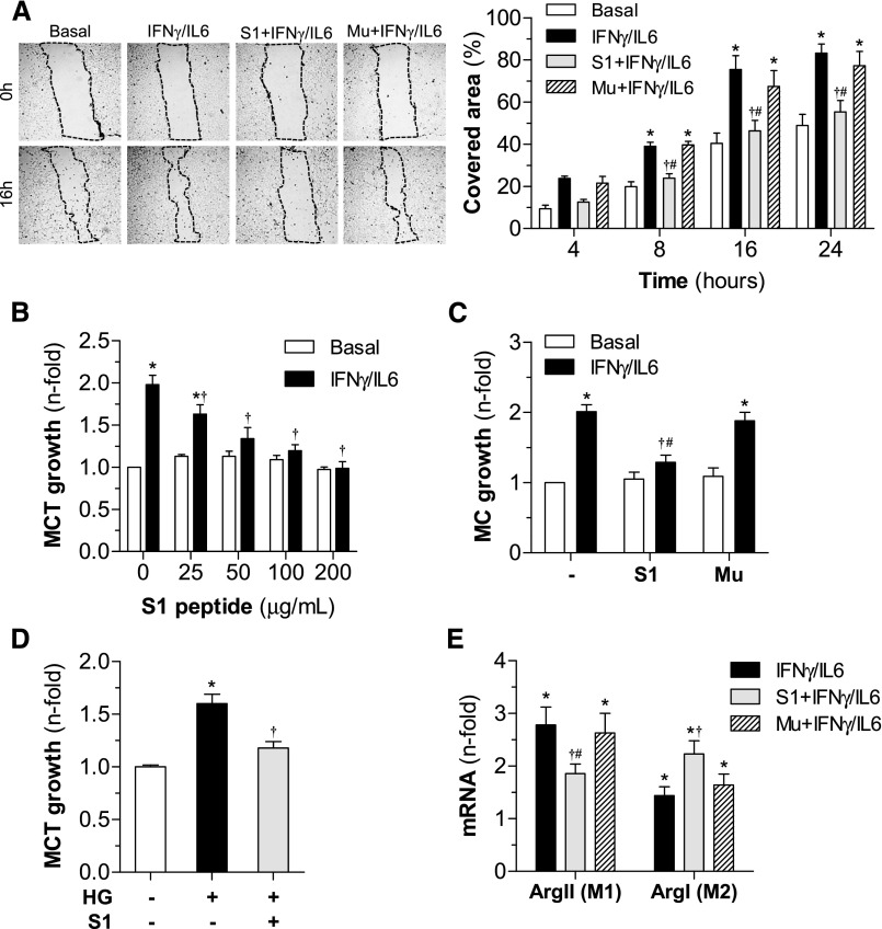
In vitro effects of SOCS1 peptide on cell migration, proliferation, and differentiation. (A) Analysis of MC migration by scratch-wound-healing assay. Representative phase-contrast images of cells migrating into the wounded area (dotted lines) at 0 hours and 16 hours of cytokine incubation in the absence or presence of peptides (S1 and Mu sequences, 100 μg/ml). The graph shows the results from quantification of covered healing areas over time. (B) Dose-dependent curves of peptides on cell viability (basal conditions) and proliferation (cytokine stimulation) in MCT (MTT assay, 48 hours). (C) Effect of peptides (100 µg/ml) on MC growth. (D) Antiproliferative effect of S1 peptide on HG-stimulated MCT. (E) Real-time PCR analysis of arginase isoforms (ArgII and ArgI) in bone marrow–derived macrophages. Data expressed as percentage or fold increases over basal conditions are mean±SEM (n=4–6 experiments). *P<0.05 versus basal; †P<0.05 versus stimulus; #P<0.05 versus Mu.

