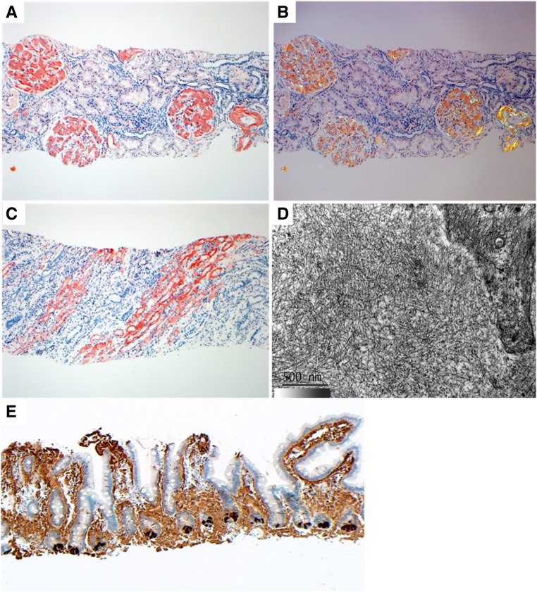
Figure 1.
Renal and GI biopsy findings. (A) Abundant glomerular mesangial, glomerular capillary wall, and arterial congophilic amyloid deposits are seen. (B) Renal CR-positive amyloid deposits display apple-green birefringence under polarized light. (C) Extensive congophilic amyloid deposits are seen in the medulla involving collecting ducts and vasa recta basement membranes. (D) On electron microscopy, amyloid deposits are composed of randomly oriented straight fibrils. (E) The intestinal mucosal amyloid deposits stain strongly for lysozyme using immunohistochemistry. Paneth cells at the bases of the glands serve as an internal positive control for lysozyme. Proteomic data confirming the diagnosis of ALys with the p.Leu102Ser abnormality from this specimen are presented in Supplemental Figure 1. Magnification, ×100 in A–C and E; ×50,000 in D.

