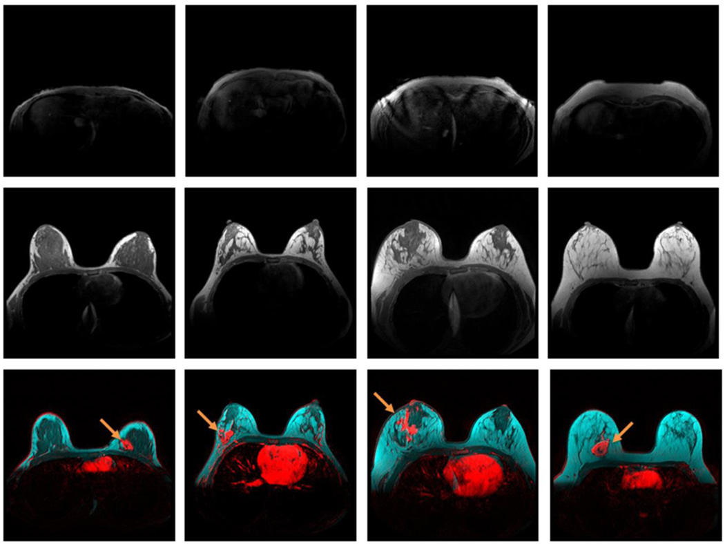
Figure 5.
Effect of body adiposity on breast adipose tissue expansion in women 31 – 32 years old. Top row: UAAL on T1-weighted pre-contrast images; center row: central breast on T1-weighted pre-contrast images; and bottom row: breast tumors (red) at the fibroglandular (dark) and adipose (cyan) tissue interface, demonstrated by breast DCE-MRI enhancement at plateau, and overlaid on T1-weighted pre-contrast images without fat saturation. Enhanced tumors are indicated by arrows. Images from left to right: a 32-year-old patient with a UAAL thickness of 2 mm, a breast density of 68.0%, and an estrogen receptor-positive IMC tumor; a 32-year-old patient with a UAAL thickness of 5 mm, a breast density of 24.5%, and an estrogen receptor-positive IMC tumor; a 31-year-old patient with a UAAL thickness of 13 mm, a breast density of 22.4%, and a triple-negative IDC tumor; and a 32-year-old patient with a UAAL thickness of 18 mm, a breast density of 5.2%, and an estrogen receptor-positive IDC tumor.

