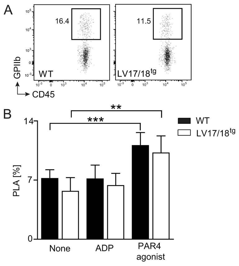
Fig. 6. Assessment of platelet-leukocyte aggregates (PLA) in WT and 2bF8 expressing mice.

GPIIb+CD45+ double positive PLA in WT and LV17/18tg mice were assessed using whole blood flow cytometry. (A) Representative dot plots of PLA pre-gated on CD45+ cells in whole blood of WT and LV17/18tg mice are shown. Values shown indicate the percentage of cell population. (B) Frequency of PLA among CD45+ events in WT (n = 7) and LV17/18tg (n = 6) blood without stimulation or upon in vitro stimulation with 20 μM ADP or 1 mM PAR4 agonist are depicted. *P< 0.05 (Student’s t-test).
