
Figure 3.
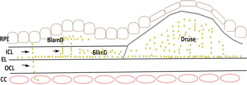
Cartoon of Bruch’s membrane deposits with AMD. Basal laminar deposits (BLamD) develop between the RPE basal lamina (RPE BL) and the RPE cell while basal linear deposits (BLinD) develop in the inner collagenous layer (ICL). A druse occupies the subRPE space and is denoted by the gray line. The yellow dots represent lipoprotein deposits, which can be released from the RPE and transit in linear streaks (arrows) toward the choriocapillaris (CC). With age-related changes to the elastic layer (EL), lipoproteins collect at the inner surface of the elastic layer (EL) and accumulate toward the RPE.