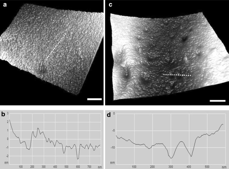
Fig. 3.
a Ultrastructure of an RBC membrane irradiated at 10 Gy. b The line profile of the image along the dashed line in (a). c Ultrastructure of RBC membrane irradiated at 15 Gy; few potholes and depressions on the cell membrane can be observed. d The line profile of the image along the dashed line in (c). Scale bar 200 nm

