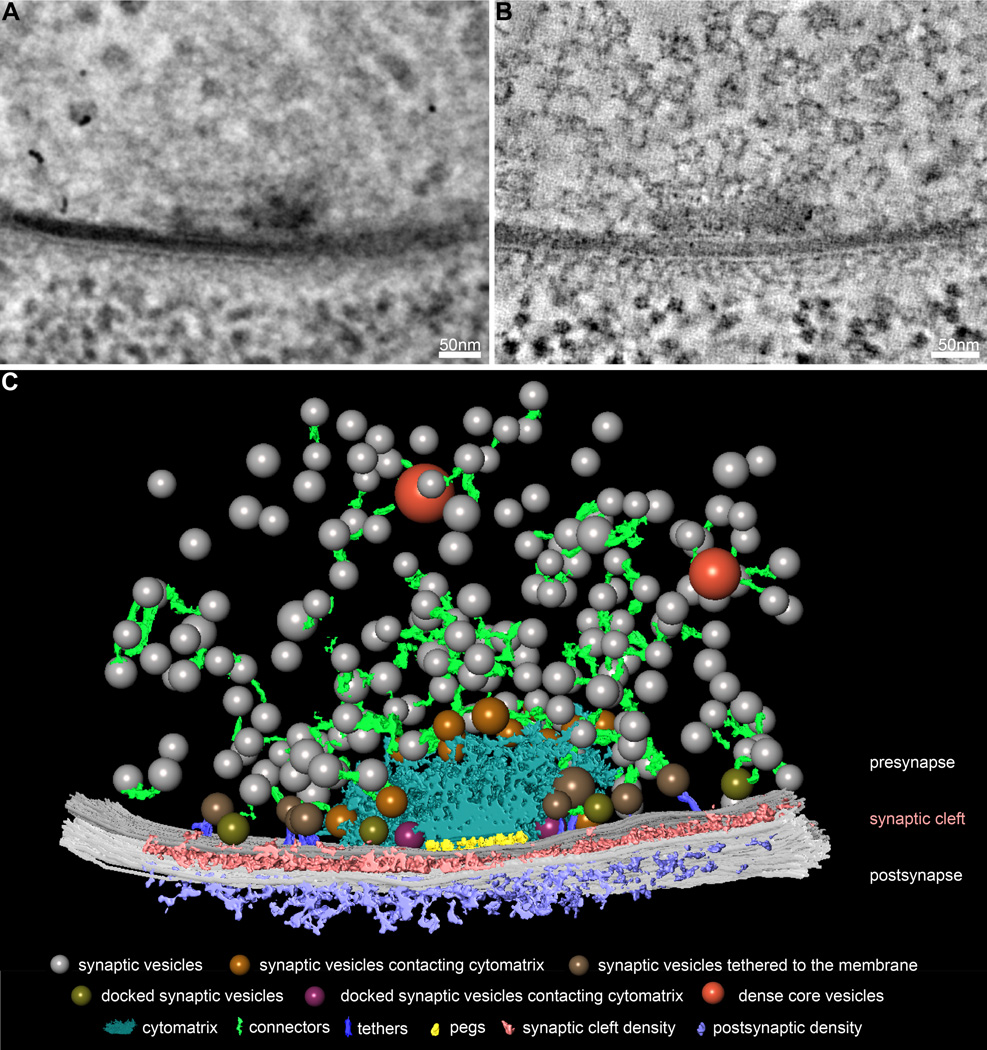
Figure 1.
Comprehensive three-dimensional reconstruction of synaptic ultrastructure at the Drosophila NMJ. (A) A representative electron micrograph of a Drosophila NMJ synapse from a central position during a 120° tilt series of a 250-nm section. (B) A representative virtual slice of the tomogram generated from the tilt series in A. (C) 3D model of segmented features of the Drosophila NMJ synapse in A and B, including synaptic vesicles (colored spheres), dense core vesicles (orange spheres), the active zone cytomatrix (blue), connectors (green), presynaptic membrane (dark gray), synaptic cleft density (pink), postsynaptic membrane (light grey), and postsynaptic density (purple). Among synaptic vesicles, we observe subsets contacting the active zone cytomatrix (gold spheres), linked to the presynaptic membrane by long tethers (brown spheres, purple filaments), directly contacting the presynaptic membrane (olive spheres), and contacting both the cytomatrix and presynaptic membrane (magenta spheres).

