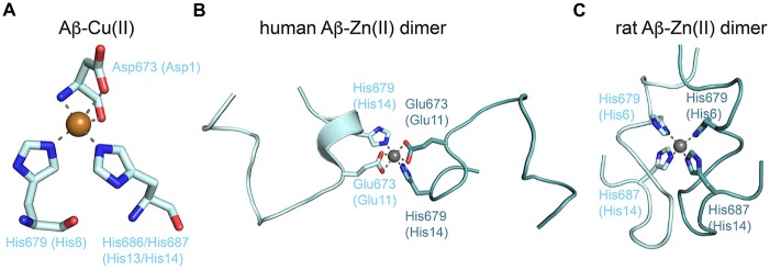
Figure 4.
Metal binding to the Aβ peptide. (A) The proposed copper (II) coordination by the N-terminus of Aβ and two histidines. The model corresponds to copper (II) coordination found in the crystal structure of GFLD-Cu(II) with the aspartate originating from a crystal contact (PDB code: 4jfn). (B) NMR structure of dimeric human Aβ (1–16) bound to zinc (II) (PDB code: 2mgt). The zinc ion mediates dimerization via coordination of equivalent residues. (C) NMR structure of dimeric rat Aβ (1–16) bound to zinc (II) (PDB code: 2li9). Dimerization via the zinc ion is different and highlights the conformational flexibility of Aβ peptides.

