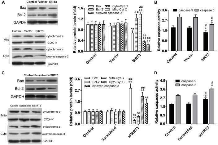
Figure 5.
SIRT3 in NSCs exerts neuroprotective effect through mitochondrial apoptotic pathway. (A) Representative western blot (left panel) and quantification (right panel) of Bax, B-cell lymphoma 2 (Bcl-2), Mito-cytochrome C (Cyt C), Cyto-Cyt C and cleaved caspase-3 protein expression in NSCs treated with empty vector or SIRT3 plasmid for 48 h. (B) Caspase-3 and caspase-9 enzyme activities were detected by the spectrophotometric method in the Control group, Vector group and SIRT3 group. (C) Representative western blot (left panel) and quantification (right panel) of Bax, Bcl-2, Cyt C and cleaved caspase-3 protein expression in NSCs transfected with scrambled RNA or SIRT3 siRNA for 48 h. (D) Caspase-3 and caspase-9 enzyme activities were evaluated by the spectrophotometric method in the Control group, Scrambled group and siSIRT3 group. GAPDH and COX-IV were used as total cellular and mitochondrial protein markers, respectively, in (A,C). Results are normalized to that of the Control group. Data are obtained in NSCs co-cultured with microglia challenged with 10 μM Aβ exposure. Data are presented as mean ± S.D. (n = 3). *P < 0.05, **P < 0.01 vs. the Control group. #P < 0.05, ##P < 0.01 vs. the Vector or Scrambled group, respectively.

